Translate

Friday, 2 August 2013

Researchers Found a switch to turn off cancer of testis and ovary


Malignant Germ Cell Tumors (GCT) are malignant tumors that are formed by immature cells that begin in the reproductive cells of the testes or ovaries. These germ cells travel into the pelvis as ovarian cells or into the scrotal sac as testicular cells. These cells metastasize to other parts of the body and most commonly spread to the lungs, liver, lymph nodes, and central nervous system. Adult germ cell tumors are usually in the testes or ovaries.

Although many patients do well after treatment, current chemotherapy treatments can have severe long-term side effects, including hearing loss and damage to the kidneys, lungs and bone marrow. For some patients, outcomes remain poor and testicular cancer continues to be a leading cause of death in young men.

Researchers from Cambridge have discovered a molecular "switch" that can turn off a highly virulent cancer of the testis and the ovary.


The scientists found that all malignant germ cell tumors contain large amounts of a protein called LIN28. This results in too little of a family of tiny regulator molecules called let-7. In turn, low levels of let-7 cause too much of numerous cancer-promoting proteins in cells.

Importantly, the cancer-promoting proteins include LIN28 itself, so there is a vicious cycle that acts as an `on` switch to promote malignancy.

The researchers have likened these changes to a `cascade effect`, extending down from the large amounts of LIN28 to affect many properties of the cancer cells.

The researchers also discovered that by reducing amounts of the protein LIN28, or by directly increasing amounts of let-7, it is possible to reverse the vicious cycle.

Both ways reduced levels of the cancer-promoting proteins and inhibited cell growth. Because the level of LIN28 itself goes down, the effects are reinforced and act as an `off` switch to reduce cancerous behaviour.

The switch effect is present in all malignant germ cell tumours, whether they occur in males or females, young or old. Such a fundamental abnormality makes an excellent new target for treating these tumours.

Thursday, 1 August 2013

A Glass of Milk After Eating Sugary Cereals May Prevent Cavities

Tooth decay is the process that results in a cavity (dental caries). It occurs when bacteria in your mouth make acids that eat away at a tooth. If not treated, tooth decay can cause pain, infection, and tooth loss.


It is known that, to avoid cavities eating high sugar and/or high acid foods (cookies, candy, soda) between meals should be avoided. According to a new research at the University of Illinois at Chicago College of Dentistry, a glass of milk after eating sugary meal may reduces plaque acid levels and may prevent damage to tooth enamel that leads to cavities.


For their study reseachers, involved 20 adults eating 20 grams of dry Froot Loops cereal, then drinking different beverages -- whole milk, 100 percent apple juice, or tap water.


They measured plaque pH or acidity with a touch microelectrode between the premolar teeth before eating, two and five minutes after eating and then two to 30 minutes after drinking a liquid.

They found that pH in plaque dropped rapidly after consuming cereal alone, and remained acidic at pH 5.83 at 30 minutes.Participants who drank milk after eating sugary cereal showed the highest pH rise, from 5.75 to 6.48 at 30 minutes. Those who drank apple juice remained at pH 5.84 at 30 minutes, while water raised the pH to 6.02.


Milk, with a pH ranging from 6.4 to 6.7, is considered to be a functional food that fights cavities because it promotes tooth remineralization and inhibits the growth of plaque.


Most consumers think that since milk is considered to be cavity-fighting, acid production by plaque bacteria can be minimized by mixing it with cereal. However, in an unpublished study in the lab, it was discovered that the combination of Froot Loops and milk became syrupy. Eating cereal combined with milk lowered plaque pH to levels similar to that obtained after rinsing with a 10 percent sugar solution.


The research is published in the July issue of the Journal of the American Dental Association.

Wednesday, 31 July 2013

Stem cells in urine easy to isolate, have potential for numerous therapies


In mammals, there are two broad types of stem cells: embryonic stem cell, which are isolated from the inner cell mass of blastocyst, and adult stem cell, which are found in various tissues. Now researchers at Wake Forest Baptist Medical Center’s Institute for Regenerative Medicine and colleagues have identified stem cells in urine that can be directed to become multiple cell types.


These cells can be obtained through a simple, non-invasive low-cost approach that avoids surgical procedures. It can be obtained just by collecting urine sample.


Researchers successfully directed stem cells from urine to become bladder-type cells, such as smooth muscle and urothelial, the cells that line the bladder. But the urine-derived cells could also form bone, cartilage, fat, skeletal muscle, nerve, and endothelial cells, which line blood vessels. The multipotency of the cells suggests their use in a variety of therapies.


To use a patient's own stem cells for therapy is considered advantageous because they do not induce immune responses or rejection. However, because tissue-specific cells are a very small sub-population of cells, they can be difficult to isolate from organs and tissues.


The team first identified the cells, which are a small subset of the many cells found in urine, in 2006. The current research builds on earlier studies by confirming the multipotency of the cells. In addition, the research found that unlike iPS cells or embryonic stem cells, the urine derived-stem cells do not form tumors when implanted in the body, indicating they may be safe for use in patients.


For their research they collected urine samples from 17 healthy individuals ranging in age from 5 to 75 years. Isolating the cells from urine involves minimal processing, according to the authors. Next, they evaluated the cells' ability to become multiple cell types.

Importantly, the cells differentiated into the three tissue layers (endoderm, ectoderm and mesoderm) that are a hallmark of true stem cells and also differentiated into the specific cell types mentioned earlier.

Next, the researchers placed cells that had been differentiated into smooth muscle and urothelial cells onto scaffolds made of pig intestine. When implanted in mice for one month, the cells formed multi-layer, tissue-like structures.

The urine-derived stem cells have markers of mesenchymal cells, which are adult stem cells from connective tissue such as bone marrow. They also have markers for pericytes, a subset of mesenchymal cells found in small blood vessels.


Researchers suspect that the cells originate from the upper urinary tract, including the kidney. Female study participants who had received kidney transplants from male donors were found to have the y chromosome in their urine-derived stem cells, suggesting the kidney as the source of the cells.


The research is published online in the journal Stem Cells.

Monday, 29 July 2013

DNA mutation breakthrough could help treat TB, cancer


A DNA mutation is any change that occurs in the DNA. These changes can be beneficial to, have some effect on, or be seriously detrimental to an organism. Each cell depends on thousands of proteins to do their jobs in the right places at the right times. Sometimes, mutations prevent one or more of these proteins from working properly. By changing a gene’s instructions for making a protein, a mutation can cause the protein to malfunction or to be missing entirely. When a mutation alters a protein that plays a critical role in the body, it can disrupt normal development or cause a medical condition.

Researchers have developed a new method to that has the capability of looking at a specific segment of DNA and point out a single mutation, which could help diagnose and treat diseases like cancer and tuberculosis.These small changes can be the root of a disease or the reason some infectious diseases resist certain antibiotics.

For example, tuberculosis -- a disease that's known to have drug-resistant strains. Its resistance to antibiotics often is due to a small number of mutations in a specific gene. If a person with tuberculosis isn't responding to treatment, it's likely because there is a mutation.


The new method has improved on previous approaches because their solution doesn`t require any complicated reactions or added enzymes, it just uses DNA. Their method is robust to changes in temperature and other environmental variables, making it well-suited for diagnostic applications in low-resource settings.

Lead author Georg Seelig, a University of Washington assistant professor of electrical engineering and of computer science and engineering along with David Zhang of Rice University and Sherry Chen, a UW doctoral student in electrical engineering, designed probes that can pick out mutations in a single base pair in a target stretch of DNA.

The probes allow researchers to look in much more detail for variations in long sequences - up to 200 base pairs - while current methods can detect mutations in stretches of up to only 20 bp.

In this method, testing probes are designed to bind with a sequence of DNA that is suspected of having a mutation. The researchers do this by creating a complimentary sequence of DNA to the double-helix strand in question. Then, they allow molecules containing both sequences to mix in a test tube in salt water, where they naturally will match up to one another if the base pairs are intact. Unlike previous technologies, the probe molecule checks both strands of the target double helix for mutations rather than just one, which explains the increased specificity.

The probe is engineered to emit a fluorescent glow if there`s a perfect match between it and the target. If it doesn`t illuminate, that means the strands didn`t match and there was in fact a mutation in the target strand of DNA.

The findings have been published online in the journal Nature Chemistry.

Saturday, 27 July 2013

Biosensor tattoo tells athletes when they're about to hit the wall


Hitting the wall is a term that athletes use to describe the condition where they physically run out of steam.This generally happens when sustained vigorous physical activity causes lactate to build up in the body. When lactate levels get too high, fatigue results. By monitoring those levels, however, athletes can ease back on the activity, allowing their lactate levels to drop before reaching that point of no return by regular blood test which is not practical during prolonged exercises.
But now a team led by the University of California, San Diego’s Prof. Joseph Wang created a flexible electrochemical biosensor tattoo that tell athelete's when they're about to hit the wall. It tells athletes when lactate levels in their body are rising, offering an early warning that the energy stores in their muscles are running low. It is applied to the skin, that temporary-transfer sensor continuously measures lactate levels in the athlete’s sweat. 

It contains an enzyme that oxidises lactate to release electrons. These electrons then generate a current through the sensor's thin and flexible electronics: the more lactate is oxidised, the larger the current. A small device attached to the tattoo measures the current to provide real-time information on lactate levels.

It was tested on 10 subjects who were working out on an exercise bike and it was able to accurately indicate the rise in their lactate levels.

In future, those readings could be transmitted by bluetooth to a cellphone, says Wang.

The technology could be used not only in the training and performance tracking of athletes, but also for soldiers and other people who are required to perform intense physical activity.

Friday, 26 July 2013

A Study Finds - Women's Height Linked to Cancer Risk


According to a new research taller postmenopausal woman have greater her risk for developing cancer. Researchers found height is linked to cancers of the breast, colon, endometrium, kidney, ovary, rectum, and thyroid, as well as to multiple myeloma and melanoma.


Furthermore, researchers were surprised at the number of cancer sites that were positively associated with height than were associated with numerous other factors linked to cancer, such as body mass index [BMI].

Senior study author Dr. Thomas Rohan, chair and professor of epidemiology and population health at Albert Einstein College of Medicine in New York City, told FoxNews.com that, they didn't find much difference in heavy or lighter women, so it’s a pretty consistent association right across the spectrum.

They studied for 12 years on around 20,928 postmenopausal women. They found that for every 10-centimeter (3.94 inches) increase in height, there was a 13 percent increase in risk of developing any cancer, for example, a woman who was 5 feet 10 inches tall would have a 13 percent higher risk for cancer than a woman who was approximately 5 feet 6 inches tall. 

Some cancer were more strongly associated with height than other. Among specific cancers, there was a 13 percent to 17 percent increase in the risk of getting melanoma and cancers of the breast, ovary, endometrium, and colon. There was a 23 percent to 29 percent increase in the risk of developing cancers of the kidney, rectum, thyroid, and blood.

Of the 19 cancers studied, none showed a negative association with height.

Researchers said that some genetic variations associated with height are also linked to cancer risk. They also said that another potential explanation may be related to environmental factors, such as childhood nutrition. Increased energy intake during childhood is thought to influence adult height and may also impact certain systems in the body. Researchers need to better understand the link exists.

Researchers will continue to explore the link between height and cancer, as they search for some of the underlying biological mechanisms that may be responsible for the correlation.

The study was published in Cancer Epidemiology, Biomarkers & Prevention, a journal of the American Association for Cancer Research.

Thursday, 25 July 2013

Researchers Developed a Way That Makes You Invisible to Mosquitoes


Several methods are used to limit exposure to mosquitoes and protect from bites when mosquitoes are unavoidable. But now researchers, including an Indian-origin scientist, have developed the world's first lightweight patch that can make people 'invisible' to pesky mosquitoes and could prove key in the battle against mosquito borne diseases.


That patch is named as 'KiteTM Mosquito patch.' It is is a 2-by-2-inch adhesive square that makes people virtually undetectable by mosquitoes for up to 48 hours. 


The patch is designed on basis of the research of University of California, Riverside in 2011, which states that mosquitoes primary way to sniff out human prey is their ability to detect carbon dioxide.

The patch, sticks to clothing, using non-toxic compounds that block mosquitoes' ability to detect carbon dioxide. It can be used by people of all ages , including infants and pregnant mothers. 

The initial research was performed in the laboratory of Anandasankar Ray, an associate professor of entomology , and was featured in the journal Nature. 

Ray's lab identified volatile odour molecules that can impair, if not completely disrupt , mosquitoes' carbon dioxide detection machinery. 

Kite's technology is the culmination of years of development work on a class of odour molecules, all of which are non-toxic compounds approved for human consumption by the US Food and Drug Administration. "The Kite Mosquito Patch isn't just another mosquito product, but a powerful alternative to most products on the market, enabling people to live normal lives with a new level of protection against contracting mosquito-borne diseases," said Michelle Brown, the chief scientist and vice president of Olfactor Laboratories.

Wednesday, 24 July 2013

A New Weapon Against Stroke


A stroke is a condition in which the brain cells suddenly die because of a lack of oxygen. This can be caused by an obstruction in the blood flow, or the rupture of an artery that feeds the brain. The patient may suddenly lose the ability to speak, there may be memory problems, or one side of the body can become paralyzed.

The treatments for stroke are currently limited, patients are treated with emergency treatments (thrombolytic drugs), which leaves survivors with severe disabilities including loss of mobility, pain, numbness, memory loss, difficulty talking and impaired cognitive ability. Many of the regenerative medicine technologies in development are targeting the post-stroke rehabilitation period for which there are currently no therapies available.

The large, collaborative study of researchers found that astrocytes -- neural cells that transport key nutrients and form the blood-brain barrier -- can protect brain tissue and reduce disability due to stroke and other ischemic brain disorders.

Astrocytes are known as housekeeping cells because of their supportive roles to neurons.They are critical to several brain functions and protect neurons from injury and death. They are not excitable cells like neurons and are easier to harness.

Researchers started their study by using a transcription factor known as Olig2 to differentiate human embryonic stem cells into astrocytes. By this approach they generated a type of astrocyte called Olig2PC-Astros and the astrocytes produced are 100 percent pure.

They then compared the effects of Olig2PC-Astros with another type of astrocyte called NPC-Astros. They conduct their research on rat in three groups. One group was transplanted with  Olig2PC-Astros, other with  NPC-Astros and third group with no transplantation.

The rats transplanted with Olig2PC-Astros experienced superior neuroprotection together with higher levels of brain-derived neurotrophic factor (BDNF), a protein associated with nerve growth and survival. The rats transplanted with NPC-Astros or that received no treatment showed much higher levels of neuronal loss.

To determine whether the astrocytes impacted behavior, the researchers used a water maze to measure the rats' learning and memory. In the maze, the rats were required to use memory rather than vision to reach a destination. When tested 14 days after transplantation, the rats receiving Olig2PC-Astros navigated the maze in significantly less time than the rats that received NPC-Astros or no treatment.

The investigators used cell culture experiments to determine whether the astrocytes could protect neurons from oxidative stress, which plays a significant role in brain injury following stroke. They exposed neurons co-cultured with both types of astrocytes to hydrogen peroxide to replicate oxidative stress. They found that, while both types of astrocytes provided protection, the Olig2PC-Astros had greater antioxidant effects. Further investigation showed that the Olig2PC-Astros had higher levels of the protein Nrf2, which increased antioxidant activity in the mouse neurons.

The investigators also investigated the genetic qualities of the newly identified astrocytes and they were genetically similar to the standard NPC-Astros.

In addition to being therapeutically helpful, the Olig2PC-Astros showed no tumor formation, remained in brain areas where they were transplanted and did not differentiate into other cell types, such as neurons.

The results could lead to stem cell treatments for many neurodegenerative diseases.

The result is published in journal Nature Communications.

Tuesday, 23 July 2013

New Key to 'Switching Off' Hypertension


Hypertension, also referred to as high blood pressure, is a condition in which the arteries have persistently elevated blood pressure. According to a medical dictionary , hypertension means "High blood pressure; transitory or sustained elevation of systemic arterial blood pressure to a level likely to induce cardiovascular damage or other adverse consequences." According to WHO, worldwide, raised blood pressure is estimated to cause 7.5 million deaths, about 12.8% of the total of all deaths. Current antihypertensive medications, control hypertension partially and most of them have serious side effects.

To minimize number of hypertension patient, researchers from University of California San Diego has designed new compounds that mimic those naturally used by the body to regulate blood pressure.


For their study researchers studied the properties of the hormone catestatin. Catestatin acts as the gatekeeper for the secretion of catecholamines -- hormones that are released into the blood during times of physical or emotional stress. Catestatin binds nicotinic acetylcholine receptors found in the nervous system, and developed a pharmacophore model of its active centers. To design a drug that mimics the action of catestatin, that would allow people to control the hormones that regulate blood pressure, they also screened a library of compounds for molecules that might match the 3D "fingerprint". Researchers created a three-dimensional model of the most important binding centers -- the pharmacophore model as in figure.


Catestatin-mimic pharmacophore model. Pharmacophore centers correspond to hydrophobic residues Leu5, Phe7, and Phe14; and positively charged residues Arg8, Arg10, and Arg15. Green circles represent hydrophobes and aromatic/hydrophobic features, while dark-blue circles represent NCN+ groups/cations/H-bond donors. Ribbon diagram and three-dimensional residue structures belong to superimposed catestatin. (Credit: Valentina Kouznetsova, UC San Diego)


After screening about 250,000 3D compound structures in the Open NCI Database, they discovered seven compounds that met the requirements, and tested those compounds in live cells to gauge their effects on catecholamines. Based on their findings, they tried one compound (TKO-10-18) on hypertensive mice, and showed that this compound produced the same anti-hypertensive effect as catestatin.

Researched is published online this month in Bioorganic & Medicinal Chemistry.

Monday, 22 July 2013

World's First Water Treatment Techniques Using Apple and Tomato Peels


Can you imagine, apple and tomato peels can be used to remove different types of pollutants in water. Till date many techniques are available for purification of water, some are - boiling, granular activated carbon filtering, reverse osmosis, use of iron in removing arsenic from water, with moringa seeds, in-situ chemical oxidation etc. Now Mr Ramakrishna Mallampati, a PhD candidate at the National University of Singapore (NUS), came up with a new purification technique. He experimented with water treatment techniques using materials that are easily available, and came up with novel ways to purify water using the peels of apples and tomatoes.


One of the most crucial problems affecting the world today is the scarcity of potable water.According to a 2007 WHO report, 1.1 billion people lack access to an improved drinking water supply. The scarcity of clean water is expected to worsen in the future due to over usage, lack of conservation methods and dwindling natural supply of clean water, even in countries with significant water resources. 


The challenge for scientists is to develop robust water purification methods that could carry out water treatment at low cost, with minimal energy consumption and using less chemicals in the process so as to reduce negative impact on the environment.In a bid to make clean water available at low cost, Mr Ramakrishna Mallampati, used materials that are easily available for water purification.


He studied the structure of the tomato peels to assess their efficiency as bio-materials to remove toxic metal ions and organic pollutants from water. In addition, factors such as the pH, nature and amount of adsorbent used for extraction were considered to establish the optimum conditions under which tomato peel could remove various pollutants from water.


He found that tomato is an efficient adsorbent for water purification. It can effectively remove different contaminants in water, including dissolved organic and inorganic chemicals, dyes and pesticides, and they can also be used in large scale applications. The results were published in the Royal Society of Chemistry journal RSC Advances in September 2012.

He also studied on apple peel for water purification as They are easily available as bio-waste from food processing industries and they are biodegradable.


He found that, similar to tomato peels, apple peels can also remove a range of dissolved water pollutants through the adsorption process. In order to enhance the ability of apple peels towards extraction of negatively charged pollutants, Mr Ramakrishna immobilized naturally occurring zirconium oxides onto the surface of apple peels. Zirconium loaded apple peels were found to be able to extract anions such as phosphate, arsenate, arsenite, and chromate ions from aqueous solutions. This method of water purification can also be used for large scale applications.


The findings are published in the American Chemical Society journal ASC Applied Materials & Interfaces in May 2013.

Sunday, 21 July 2013

Brain's 'misery' molecule discovered


Scientists have found the brain's most miserable molecule - a protein involved in all our feelings of stress, anxiety and depression - paving way for development of new drugs to control it.


Researchers used the Diamond Light Source. Diamond Light Source is the UK’s national synchrotron facility, located at the Harwell Science and Innovation Campus in Oxfordshire. By accelerating electrons to near light-speed, Diamond generates brilliant beams of light from infra-red to X-rays which are used for academic and industry research. It generates some of the world's most powerful x-ray beams that researchers used to study molecules that jut from the outer surfaces of cells in the brain's pituitary gland.


It is already known that the pituitary plays a crucial role in anxiety and depression by releasing stress chemicals into the blood. However, it was not known how the response was triggered, although a protein named CRF1 was a suspect, 'The Sunday Times' reported.

Researchers work on the protein CRF1 structure, which tells us how it works and, potentially, means they can design drugs to control it.


CRF1 is embedded in the outer membranes of pituitary cells where it looks out for stress molecules released by the hypothalamus. 


When it detects one it activates its parent cell to release hormones that, over long periods, cause anxiety and depression. 

The powerful accelerator that scientists used illuminated the molecule's entire atomic structure - including a crevice within it that could prove an ideal target for new drugs. 

The results were published in journal Nature

Eating Eggs Is Not Linked to High Cholesterol in Adolescents.


An egg is an excellent source of fats, proteins and vitamins and forms a part of healthy diet. Despite of that, eating more than two egg per week is thought to increase cholesterol as it contains around 212 milligrams of cholesterol. And, having high cholesterol levels in our blood increases our risk of heart disease.
In 1973, the American Heart Association recommended limiting egg intake to a maximum of three per week, an idea that was accepted by health experts for years.

According to the new study led by researchers at the University of Granada eating more eggs is not associated with higher serum cholesterol in adolescents, regardless of how much physical activity they do. Researchers has analysed the link between egg intake in adolescents and the main risk factors for developing cardiovascular diseases, such as lipid profile, excess body fat, insulin resistance and high blood pressure. 

According to a recent research serum cholesterol level is more affected by saturated fats and trans fats than by the amount of cholesterol in the diet. Egg has more unsaturated fats than saturated fats and only has 70 calories.


The results of this article, part of the European study HELENA involving nine countries, demonstrated that eating larger amounts of egg is neither linked to higher serum cholesterol nor to worse cardiovascular health in adolescents, regardless of their levels of physical activity.

"The conclusions, published in the journal Nutrición Hospitalaria, confirm recent studies in healthy adults that suggest that an intake of up to seven eggs a week is not associated with an increased risk of developing cardiovascular diseases," notes Soriano.

"Egg is a cheap food that is rich in very high-quality proteins, minerals, folates and B vitamins. Thus it can provide a large quantity of nutrients necessary for optimum development in adolescents," according to the researcher.

Friday, 19 July 2013

Genetic Key to Conquering Cholera


Cholera is an acute diarrheal infection caused by ingestion of food or water contaminated with the bacterium Vibrio cholerae. It is a very scary disease. Now treatments with oral rehydration therapy is available, but it is still devastating, and in extreme cases, cholera can kill in hours.

Rearchers have already discovered that there is a hereditary component that determines whether a person is susceptibility to cholera or not. According to a paper published on 4 July 2013 in the journal Science Translational Medicine. Researchers gathered genetic data from 42 families (126 individuals) of Bangladesh. They were able to find number of areas in the genome, of which some are responsible for certain immune system function, while others are related to fluid loss – that appear to be related to cholera resistance. Later tests showed genetic difference between people who had contracted the disease and those who had been exposed, but did not become ill.

A team of researchers of Harvard is now uncovering evidence of genetic changes that might also help protect some people from contracting the deadly disease.

Elinor Karlsson, the first author of the research says that, the vaccine that is available for the disease wears off after a few years, whereas people who are exposed to the disease develop a long-lasting immunity, and nobody is quite sure why that is. This research is another way of tackling that problem, and it's a way no one has come at it before.

For their study they gathered genetic data on 42 family groups – called "trios" – that included a mother, father and child. Using that data, researchers identified more than 300 areas of the genome that appeared to be under pressure due to natural selection, suggesting that genes in those regions might be adapting to deal with the threat of cholera. Which is about two percent of the genome.

To precise their finding, they turned to a process called "gene set enrichment" testing to determine whether any particular groups of genes showed up in those regions more often than others.

They found set of genes that are related to a gene called IKBKG, which plays a key role in immunity. But they also found that it was not the gene itself, but a whole group of genes that regulate IKBKG. They also found set of genes for potassium channels, which are the channels in the walls of our cells that regulate fluid loss.

Armed with that data, researchers then performed a comparative study – examining the specific genetic regions in more than 100 patients who were sick with cholera and others who had been exposed to the disease, but had not become sick. The results, showed differences between the two groups.

Researchers hope to conduct wider studies of the genetic differences between people who are susceptible and those who appear to be immune in the hope of identifying precisely which genes are involved, and the pathways involved in resistance.

Researchers discovered the largest virus ever - Pandoravirus

                                                                                                                                           
Viruses are very small in size ranging from 5 to 300 nano-meters which is not visible in a normal microscope. Researchers now have discovered a the largest virus ever - large enough to be seen in an ordinary microscope (about 1 micrometer).

The word Pandora is from Greek mythology - the first mortal woman.


The organism was initially called NLF, for “new life form”. Researchers found it in a water sample collected off the coast of Chile, where it seemed to be infecting and killing amoebae. Under a microscope, it appeared as a large, dark spot, about the size of a small bacterial cell.


They have giant-sized genome - from 1900 to 2500 genes - way more than viruses like influenza, which has 10.


The research was published in Journal science.  

                                                                                                                                                                       
Buy

New gene associated with obesity

According to WHO obesity and overweight are defined as abnormal or excessive fat accumulation that presents a risk to an individuals health. In 2005 approximately 1.6 billion adults over the of age 15+ were overweight, at least 400 million adults were obese and at least 20 million children under the age of 5 years were overweight.


In year 2007 researchers identified the first ‘obesity gene’, known as the FTO gene (Fat mass and obesity-associated protein). In may 2013, researchers has identified seven new sites on the human genome where tiny differences in the genomic structure affect the risk of obesity. Now in July 2013  researchers at Boston Children's Hospital have identified a genetic cause of severe obesity.


They conduct their research on mice. The affected gene, Mrap2 in mice, has a human counterpart (MRAP2) and appears to be involved in regulating metabolism and food consumption. They found that mice with the genetic mutation gained weight even while eating the same amount of food as their normal counterparts. Mice with gene mutation aren't burning the fat.

The protein created by the Mrap2 gene appears to facilitate signaling to a receptor in the brain called Mc4r, which helps increase metabolism and decrease appetite as part of a larger signaling chain involved in energy regulation. Fat cells produce the hormone leptin, prompting receptors in the brain to instigate production of a second hormone, αMSH. Mc4r detects this hormone with the aid of Mrap2, leading to a decrease in appetite and weight. Mutations in this signaling chain, including mutations in Mc4r, are known to increase the likelihood of obesity. 


They studied on a mice with the Mrap2 gene knocked out both overall and just in the brain. In both cases, the mice grew to about twice their normal size.Weight gain was greatest when both copies of Mrap2 were knocked out, but the mice still showed weight gain and appetite increase with one working copy of the gene. The weight gain was more pronounced in males than females.


The mice without Mrap2 didn't eat more at first, they still gained weight faster than the controls. Later, their appetites increased and they continued to gain more weight than the controls, even when held to the same diet and quantity of food. In the end, the mice carrying the new mutation must be fed about 15 percent less than normal mice to have the same amount of weight gain.


To investigate the gene in humans,researchers investigate groups of obese patients from around the world. Of 500 people with severe obesity, the researchers found only four people with the gene mutations.


While the finding suggests that these rare mutations directly cause obesity in less than 1 percent of the obese population, the researchers suspect that other mutations in the gene might occur more commonly and might interact with other mutations and environmental factors to cause more common forms of obesity.


The research was published in journal science.

Wednesday, 17 July 2013

Researcher Finds Way to Convert Blood Cells Into Autoimmune Disease Treatment

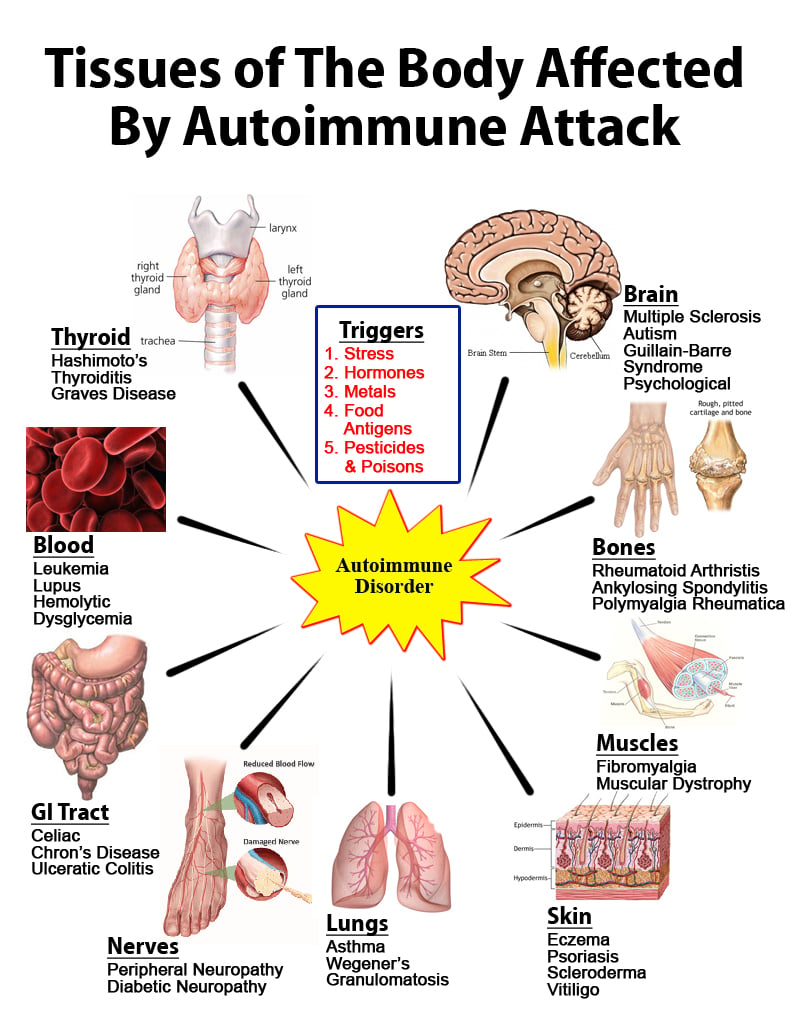
An autoimmune disorder is a condition that occurs when the immune system mistakenly attacks and destroys healthy body tissue. Normally the immune system's white blood cells help protect the body from harmful substances, called antigens.In patients with an autoimmune disorder, the immune system can't tell the difference between healthy body tissue and antigens. Examples of autoimmune disorders are rheumatoid arthritis, multiple sclerosis, addison's disease. 


Rsearchers from Purdue University discovered that Cells from one's own blood could be converted into a treatment for autoimmune diseases. Researcher has created a way to direct the differentiation of T-cells, a white blood cell that is a key player in the body's immune system.


The method involves, gathering of native T-cell from patient's blood, treated (suppressed) and then re-injected. The T cell collected is immature, from which all T-cells develop. 


Treating autoimmune diseases without compromising a patient's immune system has been a big problem in the field. But, in the research, suppressive T-cells migrate to areas of inflammation and suppress the T-cells there without significantly lowering the number of T-cells in other areas of the body where they are needed for proper immune function.

In their research they suppressed T-cell in presence of hormone progesterone. They studied on mice and found that, about 500,000 suppressive T-cells are needed to have an effect on inflammation.

This discovery and his work have been detailed in papers in the Journal of Immunology and the European Journal of Immunology. They have also filed a patent based on their work.

Tuesday, 16 July 2013

Astronomer finds new moon orbiting Neptune - 14th moon


Cape Canaveral, Florida: An astronomer studying archived images of Neptune taken by the Hubble Space Telescope has found a 14th moon orbiting the planet, NASA said on Monday.

Estimated to be about 12 miles (20 km) in diameter, the moon is located about 65,400 miles (105,251 km) from Neptune.

Astronomer Mark Showalter, with the SETI Institute in Mountain View, California, was searching Hubble images for moons inside faint ring fragments circling Neptune when he decided to run his analysis program on a broader part of the sky.
 Read more...

Missing Brain Enzyme Leads to Abnormal Levels of Fear in Mice, New Research Reveals

Fear is a vital response to physical and emotional danger—if we didn't feel it, we couldn't protect ourselves from legitimate threats. But new research from neuroscientists and molecular biologists at University of Southern California shows that a missing brain protein may be the culprit in cases of severe over-worry, where the fear perseveres even when there's nothing of which to be afraid.

Researchers examined mice without the enzymes monoamine oxidase A and B (MAO A/B), which sit next to each other in our genetic code as well as on that of mice. In their prior research they found an association between deficiencies of these enzymes in humans and developmental disabilities along the autism spectrum such as clinical perseverance (the inability to change or modulate actions along with social context).

They compare mice without MAO A/B with their wild-type litter-mates. both type of mice were put in a new, neutral environment and given a mild electric shock. All mice showed learned fear the next time they were tested in the same environment but with the MAO A/B knockout mice displays a greater degree of fear.

But while wild mice continued to explore other new environments freely after the trauma, mice without the MAO A/B enzymes generalized their phobia to other contexts — their fear spilled over onto places where they should have no reason to be afraid.

The mice without MAO A and MAO B also learned eye-blink conditioning much more quickly than wild mice, which has also been noted in autistic patients but not in mice missing only one of these enzymes.

Researchers found that the mice without MAO A/B did not display any differences in learning for spatial skills and object recognition, but in their ability to learn an emotional event, the [MAO A/B knockout mice] are very different than wild types.

When both enzymes are missing, it significantly increases the levels of neurotransmitters, which causes developmental changes, which leads to differential expression of receptors that are very important for synaptic plasticity — a measure of learning — and to behavior that is quite similar to what we see along the autism spectrum.

Monday, 15 July 2013

Your eyes offer a peek into your brain's health

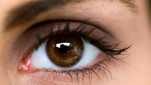
Eyes are organs that detect light and convert it into electro-chemical impulses in neurons. But the small vessels behind eyes could also reveal how healthy your brain is, a new study has revealed. 

Scientists have found that people with wider veins scored worse on IQ tests in middle age, Fox News reported.

The study`s lead author, Idan Shalev, said that factors like smoking, diabetes, or socioeconomic status couldn`t be alone blame for the poor scores.

Shalev said that because eye vessels are developed from the same cells that brain vessels are developed from, they may reflect its condition. The study has found that the health of your eyes could indicate brain health at a much earlier age.

Shalev said that even if you're blessed with 20/20 vision, retinal imaging- a fancy term for the photo eye docs take of your eyes- does far more than test vision and It could be the easiest way yet to check in on your brain.

Antiviral Enzyme Contributes to Several Forms of Cancer

Antiviral enzymes are enzymes that cells normally use to fight viruses. It is also involved in keeping cancer cells alive, researchers at UT Southwestern Medical Center have discovered on October 2006.

In February 2013 it was founded that APOBEC3B is an enzymatic source of mutation in breast cancer. Now researchers at the University of Minnesota have discovered that a human antiviral enzyme causes DNA mutations that lead to several forms of cancer.

Researchers analysed tumor samples from 19 different types of cancer for the presence of APOBEC3B and 10 related proteins. Their result showed that APOBEC3B alone was significantly elevated in six types (bladder, cervix, two forms of lung cancer, head & neck, and breast). Levels of the enzyme, is present in low levels in most healthy tissues.

They also find that the mutational signature of APOBEC3B is a close match to the actual mutation pattern in these cancers.

Findings from both studies are counter intuitive because the enzyme, which is produced by the immune system, is supposed to protect cells from HIV and other viruses, not harm our own genomic DNA.

While it's well known that sunlight and chemical carcinogens can mutate DNA, and that mutations are essential for cancer to develop, Harris is the first to discover that this human enzyme is a major cause mutation in cancer. Reuben Harris, a professor of biochemistry, molecular biology and biophysics based in the College of Biological Sciences and is also a member of the Masonic Cancer Center, University of Minnesota.

Harris believes that APOBEC3B is a biological "double-edged sword" that protects some cells from viruses such as HIV and produces mutations that give rise to cancer in others.

Harris hopes to find a way to block APOBEC3B from mutating DNA, just as sunscreen blocks mutations that lead to melanoma. It's also possible that a simple test for APOBEC3B could be used to detect cancer earlier. 

The study was published in the journal Nature Genetics.

Sunday, 14 July 2013

Gene Therapy Using Lentivirus to Treat Wiskott-Aldrich Syndrome Promising

Wiskott–Aldrich syndrome (WAS) is a rare X-linked recessive disease. This condition primarily affects males. It is characterized by abnormal immune system function (immune deficiency) and a reduced ability to form blood clots. This condition primarily affects males. 

It is caused by a mutation in a gene that encodes the protein WASP. This protein is found in all blood cells. WASP is involved in relaying signals from the surface of blood cells to the actin cytoskeleton, which is a network of fibers that make up the cell's structural framework. WASP signaling activates the cell when it is needed and triggers its movement and attachment to other cells and tissues (adhesion). In white blood cells, this signaling allows the actin cytoskeleton to establish the interaction between cells and the foreign invaders that they target (immune synapse).

Treatment of Wiskott–Aldrich syndrome is currently based on correcting symptoms. Management can accomplished through 'Cord blood & Bone marrow transplant' . It can be curative for some patients, those who have a strongly matching donor.

An alternative is to obtain blood stem cells from patient, and, in the laboratory, use gene therapy involving a form of retrovirus to take the normal gene into the cells to correct the defect. The patients are then given back the genetically changed blood cells back. This approach has been successful . However, over the long term, patients developed blood cancers.

Researchers now have developed a new gene therapy approach to the treatment of Wiskott-Aldrich Syndrome. They used a partially inactivated lentivirus to take the normal gene into the cell, while reducing the risk of cancer by inserting gene next to a cancer-promoting gene.

For their research they took the patients own blood stem cells in the laboratory, and used the lentiviral vector combined with the normal WASP gene to correct the genetic defect in the blood.

After a special treatment to eliminate their defective immune system, they received their own blood cells that had been altered to contain the normal WASP gene.

The research was published in Science Express.

Saturday, 13 July 2013

Interspecies Transplant Works in First Step for New Diabetes Therapy

Diabetes means increase in blood glucose or blood sugar levels. Insulin is a hormone that is important for metabolism and utilization of energy from the ingested nutrients - especially glucose. Without insulin, too much glucose stays in blood.

Insulin is synthesized in significant quantities only in beta cells of islets of Langerhan in the pancreas. It is secreted primarily in response to elevated blood concentrations of glucose. Insulin thus can regulate blood glucose and the body senses and responds to rise in blood glucose by secreting insulin. In type 1 diabetes, pancreas does not make or make in less quantity insulin.

Treatment of diabetes is a lifelong commitment to: Taking insulin, exercising regularly and maintaining a healthy weight, eating healthy foods nad monitoring blood sugar level. For people with hard-to-control type 1 diabetes, a transplant of insulin-producing islets from a deceased donor is one important way to control their chronic disease, in which their bodies do not produce insulin. However, there is a severe shortage of islet cells from deceased donors. Many patients on waiting lists don't receive the transplant or suffer damage to their heart, nerves, eyes and kidneys while they wait.

Scientists have been trying to transplant cells that produce insulin from other species to humans, but concerns about controlling rejection of transplants from a different species have made that approach insurmountable until now. Scientists from Northwestern Medicine® have successfully transplanted islets, from one species to another. And the islets survived without immunosuppressive drugs.

Scientists persuaded the immune systems of mice to recognize rat islets as their own and not reject them. Notably, the method did not require the long-term use of drugs to suppress the immune system, which have serious side effects. The islets lived and produced insulin in the mice for at least 300 days, which is as long as scientists followed the mice.

In their study they, removed the rat splenocytes (a type of white blood cell located in the spleen) and treated with a chemical that caused their deaths. Next, the dead splenocytes were injected into the mice. The cells entered the spleen and liver and were mopped up by scavenger cells. The scavengers processed the splenocytes and presented fragments of them on their cell surface, triggering a reaction that told the T cells to accept the subsequently transplanted rat islets and not attack them.

But rejection was still a threat. A unique challenge of an interspecies transplant is controlling the B cells, immune cells that are major producers of antibodies. Initially, when scientists transplanted the rat islets into the mice, the mouse immune system started producing antibodies against the rat cells causing rejection.

To solve the problem, scientist realized that they needed to kill off the B-cells at the same time they injected the donor islets into the mice. Thus, they gave the mice B-cell depleting antibodies -- already used in a clinical setting in human transplants. When the B-cells naturally returned after the transplant, they no longer attacked the rat islets.

Their ultimate goal is to be able to transplant pig islets into humans, but the barrier from rats to mice is probably lower than from pigs to humans. Now they are trying to figure out why the B-cells are different when they come back.

The study is published in the journal Diabetes.

Latest News - an efficient system to coat tiny objects

University of Melbourne researchers have developed an efficient system to coat tiny objects, such as bacterial cells, with thin films that assemble themselves which could have important implications for drug delivery as well as bio-medical and environmental applications.

The research is published in the journal Science, Professor Frank Caruso from the Department of Chemical and Biomolecular Engineering at The University of Melbourne and his team have developed a new strategy to coat microscopic materials. 
Read more...

Thursday, 11 July 2013

A novel way of killing cancer cells

Cancer cells is a cell that divides and reproduces abnormally with uncontrolled growth. Cancer cells can spread to other parts of the body through the blood and lymph systems. In cancer cells the normal control systems that prevent cell overgrowth and the invasion of other tissues are disabled. They have a much higher demand for nutrients and oxygen than a normal cell.

Till date several treatments are available for treatment of cancer - surgery, chemotherapy, radiation therapy, biologic or targeted therapy. Chemotherapy and radiotherapy cause damage to healthy cells and other treatments are usually only effective for individual types of cancer. But researchers from University of Southampton  in collaboration with colleagues at the BC Cancer Agency Research Centre have discovered a novel method to kill cancer cells, which leaves the body's healthy cells undamaged.


They have discovered a cellular component, eEF2K, plays a critical role in allowing cancer cells to survive nutrient starvation, whilst normal, healthy cells do not usually require eEF2K in order to survive. They report that by blocking the function of eEF2K, cancer cells can be killed, without harming normal, healthy cells in the process.

eEF2K gene encodes a highly conserved protein kinase in the calmodulin-mediated signaling pathway that links activation of cell surface receptors to cell division. This kinase is involved in the regulation of protein synthesis. It phosphorylates eukaryotic elongation factor 2 (EEF2) and thus inhibits the EEF2 function.


They are now working with other labs, including pharmaceutical companies, to develop and test drugs that block eEF2K, which could potentially be used to treat cancer in the future.

Professor Proud is also researching the origins of cancer. He says that protein synthesis -- the creation of proteins within cells -is a fundamental process that enables cells to grow, divide and function. If it goes wrong, it can contribute to the development of cancer. They are interested in how defects in this process can cause cancers and other diseases.

The research is published in journal cell.

Key Protein Revealed as Trigger for Stem Cell Development

Stem cells are mother cells that have the potential to become any type of cell in the body. One of the main characteristics of stem cells is their ability to self-renew or multiply while maintaining the potential to develop into other types of cells. This ability of stem cells have made researchers to identify how undifferentiated stem cells become the differentiated cells that form the tissues and organs. Scientists know that turning genes on and off is central to this process. 


Researchers from the Medical Research Council Centre for Regenerative Medicine at the University of Edinburgh have identified a protein - Tcf15, that kick-starts the process by which stem cells can develop into different cells in the body. 

They conduct their studies on embryonic stem cells of mice. They have also developed a technique enabling them to highlight the presence of the key protein -- Tcf15 -- in the cells. By the technique researchers can identify which cells have the protein and watch how it affects stem cells in real time to gain a better understanding of how it works.

Their discovery could help scientists improve techniques enabling them to turn stem cells into other cell types in the laboratory. These could then be used to test drugs or help create therapies for degenerative conditions. 

Researchers pinpointed the protein by looking at how some stem cells are naturally prevented from specializing into other cell types.

They found two sets of proteins, one of which binds to the other blocking them from carrying out their various functions.

They were then able to screen the blocked proteins to find out which ones would enable stem cells to differentiate.

The research is published in the journal Cell Reports.